The right lung is shorter broader and has a greater volume than the left lung. Indentation on the medial side of each lung where the bronchus pulmonary arteries and nerves enter the lung and the pulmonary veins exit.

Lung Wikipedia
Primary bronchus and the pulmonary artery and veins.

. Hilus or hilum Indentation on mediastinal medial surface Place where blood vessels bronchi lymph vessel and nerves enter and exit the lung Root of the lung Above structures attaching lung to mediastinum Main ones. The alveoli of the lungs are made of alveolar type 1 cells which are what type of tissue. Collectively these structures attach the lung to the mediastinum and.
They are suspended from the mediastinum by the lung root a collection of structures entering and leaving the lungs. Blood supply of lungs 9. Structure that contains the vocal cords and is a passageway.
Located in the neck inferior to the pharynx. On the medial mediastinal surface of each lung is an indentation the hilum through which blood vessels bronchi lymphatic vessels and nerves enter and exit the lung. Collectively these structures attach the lung to the mediastinum and are called the root of the lung.
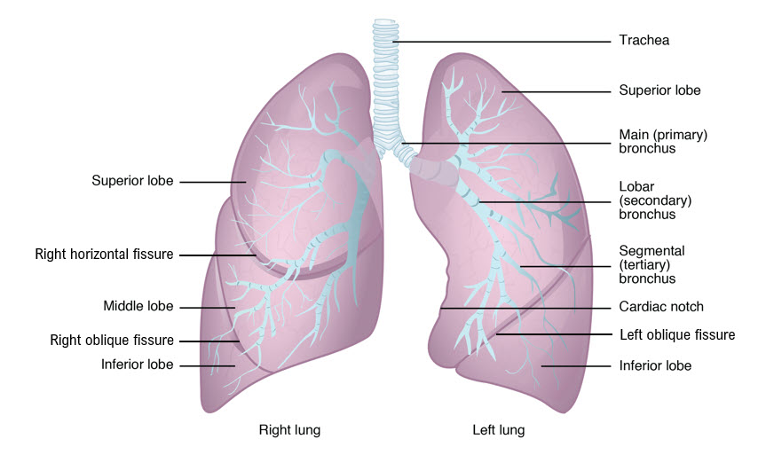
As a result the medial border of the left lung has a deep indentation called cardiac notch. Medial view R lung Medial view of L lung. This is the region where the major blood vessels bronchi and nerves enter and leave the lung.
The broad concave inferior portion or base of each lung rests on the superior surface of the diaphragm. The left lung is longer and narrower than the right lung. The left lung has a cardiac notch which is.
The hilum of the lung is the wedge-shaped area on the central portion of each lung located on the medial middle aspect of each lung. Indentation on mediastinal medial surface Place where blood vessels bronchi lymph vessel and nerves enter and exit the lung Root of the lung Above structures attaching lung to mediastinum Main ones. The three-lobed right lung is larger because the two- lobed left lungs space has to accommodate the heart as well.
The hilar region is where the bronchi arteries veins and nerves enter and exit the lungs. Lymphatics of Lungs 10. Together these structures form the root of the lung.
Nerve supply of Lungs 11. It has an indentation called the cardiac notch on its medial surface for the apex of the heart. The anterior aspect of the medial surface is referred to as the anterior mediastinal part while the dorsal half is known as the posterior vertebral part.
The surface of the lung is divided in lobules by connective tissue septa walls that are extensions of the connective tissue of the visceral pleura. On the medial mediastinal surface of each lung is an indentation the hilum through which blood vessels bronchi lymphatic vessels and nerves enter and exit the lung. The lungs are not symmetrical.
Indentation on the medial side of each lung Larynx. Gross Anatomy of Lungs 2. On the medial edge of the left lung is a deep indentation called the ___This notch accommodate the left facing apex of the heart Hilum The medial surface of each lung has a slit shaped area called the ___where vessels and bronchi enter and exit.
It is divided into three lobes and each lobe is supplied by one of the secondary bronchi. Alveolus hollow sphere of cells in the lungs where oxygen and carbon dioxide are exchanged. Fissures and Lobes of Lungs 5.
This indentation provides room for the apex of the heart. The lungs lie either side of the mediastinum within the thoracic cavity. Anatomical Position and Relations.
The indentation on the medial side of each lung is called the _____ hilus. Option 1 restricts the movement of the anterior and posterior walls of the lung and option 2 restricts the expansion of the lateral wall of the lung. A prominent indentation called the cardiac notch is also present along the mediastinal surface of the left lung.
Pulmonary and systemic blood vessels and bronchi lymphatic vessels and nerves enter and leave the lung at this point. This is where the vocal cords are located. Pulmonary artery and veins and main bronchus 10 Medial view R lung Medial view of L lung.
Surfaces and Borders of Lungs 3. Histopathology of Alveoli 7. The left lung has two lobes.
Like the other lung surfaces the medial surface has numerous indentations left by the adjacent structures that make an impression on the surface. The _____ _____ and the _____ ___ enter the lung. Pulmonary artery and veins and main bronchus.
Indentation on the medial side of each lung where the bronchus pulmonary arteries and nerves enter the lung and the pulmonary veins exit. Indentation on the medial aspect of each lung. The medial surfaces of both lungs lie in.
Hilo- hilum indentation in an organ larynx. Each lungs mediastinal surface has an indentation known as the hilum. Twitching under left rib cage.
Expands ad contracts with each breath. Hilum and Root of Lungs 4. Each lung is surrounded by a pleural cavity which is formed by the visceral and parietal pleura.
On The Medial Surface Of Each Lung Is An Indented

Lungs Anatomy Organs In Thorax
![]()
Lung Anatomy Blood Supply Innervation Functions Kenhub
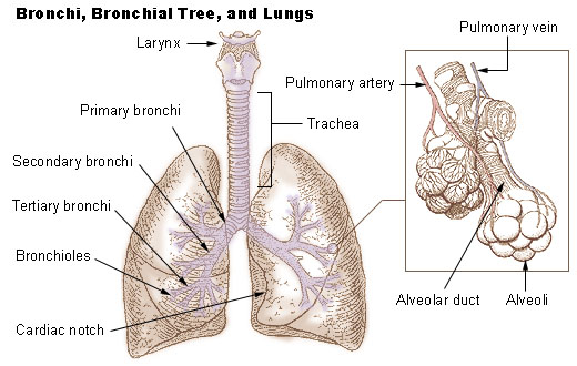
Seer Training Bronchi Bronchial Tree Lungs
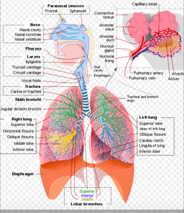
Respiratory System Physiopedia
20 3 The Lungs Medicine Libretexts

Medial Surface Of The Lungs The Medial Mediastinal Surfaces Of The Right And Left Lungs Present Concave Mirror Images Of The Ri Lunges Lung Anatomy Surface
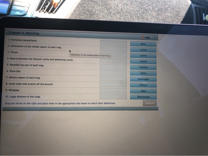
Solved Le Chapter 4 Matching Help X 1 Pulmonary Parenchyma Chegg Com
0 comments
Post a Comment